Alluce rigido: intervistato dalla rivista STARBENE
18 Marzo 2023Since 2016 I have been a member and co-founder of PBS. PBS is the acronym of Percutaneous Bianchi System and represents the work philosophy of Dr Bianchi’s entire medical team. The purpose of our work is aimed at correction of the acquired defects of the forefoot.
Over the years, our team have continually searched for less invasive, yet increasingly effective solutions to treat conditions of the forefoot, especially bunions. We have developed and shared a surgical approach, inspired by keyhole techniques and which we have adapted to the specific needs of the forefoot and bunions. By continually striving to keep this approach as natural as possible, Our team can resolve bunion problems with rapid, mini-invasive surgery without using fixative devices (screws, plates or wires). The fractures stabilise naturally, thanks to post-surgery bandaging and other innovations perfected in recent years to allow patients to walk immediately and rapidly recover the use of the foot and, therefore, their psycho-physical wellbeing. A thorough knowledge of how the foot works and an analysis of the numerous case studies have enabled the PBS team to refine the techniques and surgical procedures, to develop personalised instruments and draw up a step-by-step procedure to treat these conditions. This begins with a medical assessment of the patient’s condition and continues right up to the complete recovery of normal use. Perfection of the technique and procedures has enabled the PBS team to use keyhole surgery to treat bunions, hammer toe, metatarsalgia and the fifth metatarsus varus.
At the heart of the PBS philosophy is its continual, on-going research into foot conditions. It aims to provide treatment, which complies with the laws of biomechanics and the physiology of the foot. PBS is, therefore, a place where all medical specialists (surgeons, physiatrists, physiotherapists, podiatrists and orthopaedic technicians) can collaborate and exchange ideas to encourage continual, on-going innovation. The PBS philosophy attaches great importance to the patient’s wellbeing. This has meant the team has been able to systematically develop a series of steps to treat bunions and conditions of the forefoot over the years. The professionalism and the competence of the PBS team accompany the patient from the initial consultation. The procedure is not just surgery, which we suggest only when there are no alternative solutions. It often favours conservative therapies, which are able to prevent the condition from getting any worse.
Conditions
Bunion (Hallux Valgus)


Fig. 1 a Fig. 1 b
Numerous people, especially women, suffer from the foot condition known as a bunion (Fig. 1 a). It is crippling condition, the causes of which are not completely understood, even though it is often hereditary and can become worse, if you wear the wrong type of shoes. In the most serious cases, surgery is required to correct Hallux Valgus. In recent years, the trend has been to use mini-invasive, keyhole techniques to operate on patients which, in addition to giving excellent results, allow much shorter recovery times compared to traditional surgical techniques. The use of this technique for purely aesthetic reasons is, however, totally excluded.
The causes can, therefore, be genetic (idiopathic Valgus) or secondary, i.e. linked to other conditions (calcaneal Valgus and pronation of the talus joint – Fig. 1 b) or to autoimmune and degenerative conditions, which cause structural alterations of the foot.
Footwear, however, does not directly cause Hallux Valgus, although very high heels or very narrow toe caps can worsen any inflammation already present in the area and aggravate an existing bunion, due to the mechanical conflict involved. The deformation of the big toe towards the other toes of the foot is the principal symptom of the condition.
It is often very painful, especially if pressed or if weight is loaded on the part of the foot affected by the bunion. The appearance of a bony protuberance or “bunion” is the visible indication of the condition and it can worsen if the shoe compresses it. The affected toes may also have many calluses and red, hardened skin with reactive bursitis, which can also become infected or ulcerated.
If the condition is serious, it can cause a change in the shape of the foot, highlighted by a progressive deformation of the shoes, which can become increasingly uncomfortable and difficult to wear. These changes can also cause difficulty in walking, due to the important role the big toe plays in pushing the body forward and balancing your weight.
In order to slow the process of worsening of the bunion it is extremely important to treat the condition as soon as possible. The principal non-surgical treatments consist of the use of orthotics, sole-inserts and painkillers accompanied by the use of broad-soled comfortable shoes with low heels. The moment these so-called “conservative” do not bring any further benefits, operation will then become necessary.
Metatarsalgia

Fig. 2
Metatarsalgia is a condition characterised by the inflammation of the plantar forefoot, (Fig. 2) which causes pain in the metatarsal bones.
In general, this condition is caused by several factors acting together. These include: high heels, overweight/obesity, serious anatomic deformities (cavus foot, hammer toe, Hallux Valgus), rheumatoid arthritis, gout, very intense training or physical activity involving the part affected by the condition (e.g. running, tennis, football, etc.), stress fractures, Morton’s Neuroma (which affects the nerves between the various metatarsals), ankle or inflamed Achilles tendon, osteochondrosis (process of necrosis of the second metatarsal of the foot) and diabetes. After a careful clinical analysis, the doctor will decide whether it is better to treat the metatarsalgia with a conservative therapy (in the majority of cases) or whether surgery is necessary (only in very serious cases).
Non-surgical therapy consists mainly of small changes in your lifestyle, which can help to ease or even make the symptoms disappear. These include resting the foot affected by metatarsalgia as much as possible, applying ice to help reduce the inflammation, raising the limb to reduce load induced stress, wearing suitable shoes, if possible using anti-shock insoles or orthotics for cavus foot. If the pain is more severe, you should take painkillers and anti-inflammatory medication. If you are overweight, then you will be advised to lose weight and to do stress-free sport (e.g. swimming). Finally, for conditions such as diabetes, rheumatoid arthritis or gout, you are advised to keep them under control, as this also helps mitigate the consequences of metatarsalgia.
Surgical treatment of the condition is a solution, which is generally considered only for particularly serious cases (serious deformities of the foot or Morton’s Neuroma), which cannot be treated using the other solutions indicated.
Hammer toe

Fig. 3
Hammer toe appears as hyperflexion of one or more toes of the foot at the intermediate interphalangeal joint ( Fig. 3) his anomalous flexion leads to the formation of a painful callous with dorsal bursitis, which may ulcerate on contact with footwear. This deformity can be either fixed or flexible and can arise in young people or as the consequence of a trauma.
This condition is often associated with bunions. In this case, the hammer toe is hypertensive on the first phalange and flexed on the second. Hammer toe can be caused by arthrosis of the toes in individuals who suffer from cavus foot, traverse flatfoot of the forefoot, or who wear narrow, tight shoes and/or particularly rigid shoes.
Physical therapy can bring great relief and the use of external supports (orthoses) helps to improve the position of the toe and to reduce the discomfort caused by shoes rubbing.
Over time however, hammer toe may worsen and stiffen due to the retraction of the joint capsules and the doctor may diagnose the need for surgery. Recommended surgery involves the use of a mini-invasive percutaneous (keyhole) technique, as it does not use pins, metal wires or other fixation devices. It also encourages a quicker recovery.
Varus of the 5th Metatarsal

Fig. 4
The varus fifth toe appears as a dorso-lateral bowing of the head of the 5th metatarsal. It is usually accompanied by pain, swelling and the varus deviation of the 5th metatarsal (Fig. 4), and can also involve overlapping of the 4th metatarsal. This condition is congenital and is mostly linked to a shorter extensor tendon of the 5th toe.
One of the main reasons why this condition appears is
use of tight shoes (or narrowed toes or high heels). However, it can also be due not only to hereditary factors, but also to rheumatic or neurological disorders or to another anomaly of the foot (secondary causes).
Conservative treatment may include bunion padding, physical therapy or physiotherapy or the use of silicon protectors (orthoses).
If these solutions prove to be ineffective, then you need corrective surgery which, in recent years, mainly uses the mini-invasive, keyhole technique performed under local anaesthesia.
SURGICAL TREATMENT
The surgical treatment can be carried out in Day Hospital. On the same day of the surgery, you will be given post-surgical footwear and padded bandaging, so you can place your feet on the ground and walk normally without using crutches. The steps in the PBS Procedure are:
1. The initial consultation
The PBS procedure begins with the initial consultation to establish the extent of the condition and determine whether the patient can be treated with medication or requires surgery. The patient should come to this initial consultation with a bilateral foot X-Ray in a standing position.
2. Preparation for surgery and hospitalisation
The patient will be placed on the surgery waiting list at the clinic with the doctor of his choice and will be contacted by the clinic to undergo preoperative tests.
3. Surgery


Fig. 5 Fig. 6
Anaesthesia (Fig. 5) and surgery with PBS instruments (Fig. 6) without fixation devices (screws, wires, plates, pins). Assessment of alignment, dressing and specific PBS bandaging. Anaesthesia of the foot can be achieved using many techniques. Keyhole surgery is unusual as it requires the immediate mobility of the limb, and needs the patient to be pain-free after surgery, in order to achieve the ideal ankle block. The ankle block is a selective, local anaesthesia, which only suppresses the sensation of pain (whereas sensitivity to touch, for example remains unaltered). Together with a tranquiliser, administered before carrying out the block, makes it acceptable to most of the patients.Surgical Operation for Hallux Valgus and other pathologies with dental burrs are used to correct the deformity and modify the bones of the forefoot. These are inserted through small, approximately 2-3-millimeters incisions in the skin. The surgeon guides the burr using a special radiological device to help him “see” the bones and the burrs. The most important innovation of the technique is that it does not use fixative devices (screws or wires). This allows the fractures to heal according to the “needs” of the foot, a foot which will be able to walk immediately after surgery.
4. Post-surgery
At the end of surgery, the surgeon provides a personalised plan for the post-surgery procedure, depending on the complexity of the surgical procedure; the foot is bandaged and taped using a technique specifically developed by the medical team. After surgery, you will be able to wear an orthopaedic shoe (Fig. 7) and take your first steps. At home, you must gradually and progressively increase walking, alternating walks and rest.

Fig. 7
5. Monitoring
After 21/30 days, you should return to the clinic for a check-up. The time varies depending on the condition operated on. The medical staff will check the progress of the healing process and provide personalised procedures: exercises and possibly medication, normal shoes (Fig. 8) to help reach a successful conclusion. Further check-ups may be required in the 3 months after surgery.

Fig. 8
6. X-ray Check-up
90 days after surgery, you will have a routine check-up with the surgeon to assess how calcification, consolidation and alignment of the bones of the foot are progressing. You should bring your X-rays (Fig. 9) of the orthostatic load to this check-up (fig. 10 a before surgery, Fig. 10 b 90 days after surgery).
 .
. 
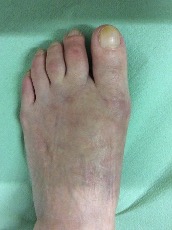
Fig. 9 Fig. 10 a Fig. 10 b